White blood cell pattern tool
Hemra shows which blood cell an image resembles.
Upload a microscope image of a white blood cell. Hemra scores it against four learned cell types, privately in your browser.
- Runs on your device
- Nothing is uploaded
- Four cell types
- Not a diagnosis
- 1Upload
- 2Confirm
- 3Results
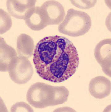
These are the model's similarity scores: how closely the image resembles each learned pattern. They are not a diagnosis.
How it works
- 1Upload
Choose a stained blood smear image with one white blood cell near the centre.
- 2Compare
A neural network in your browser compares it with patterns learned from thousands of cell images labelled by clinical pathologists [1].
- 3Read
You get a score for each cell type. On 960 held-out images from the same dataset, the top score matched the pathologists' label 97.5% of the time (see results). Expect lower accuracy on images from other labs or stains.
The four cell types

Built by Teddy Aaron, a student founder in Greenwich, Connecticut. About Teddy →
Private by design
Your image is analyzed in your browser and is never uploaded or stored. The page loads its code and fonts from public CDNs (jsDelivr and Google Fonts). Hemra has no accounts, cookies or analytics.
Research only
Hemra is an educational tool, not a medical device. It learned only from normal cells of people without infection, blood disease or cancer [1], so it cannot detect leukemia or any other condition. Its scores are not the chance that anyone is ill.
Sources
- Acevedo A, Merino A, Alférez S, Molina Á, Boldú L, Rodellar J. A dataset of microscopic peripheral blood cell images for development of automatic recognition systems. Data in Brief. 2020;30:105474. doi:10.1016/j.dib.2020.105474
- Acevedo A, et al. Peripheral blood cell image dataset (Hospital Clinic of Barcelona; May Grünwald-Giemsa stain; CellaVision DM96). Mendeley Data, v1. doi:10.17632/snkd93bnjr.1. Licensed CC BY 4.0.
- MedlinePlus, U.S. National Library of Medicine. Blood Differential.
- Haematologica Atlas of Hematologic Cytology. Normal peripheral blood cells.
All cell images, including the samples on this page, come from the dataset in [2], used under CC BY 4.0, via the copy at Bbennett21/White-Blood-Cell-Classification. Changes: Hemra uses a four-class subset of the images, cropped and resized. The dataset's authors have not endorsed Hemra.